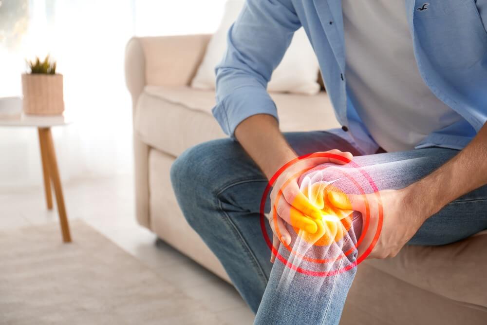
What is knee bursitis, and how physiotherapy can help?

Image source: Shutterstock
What is a bursa?
During joint movement, tendons, ligaments and muscles glide over the bones. To reduce the friction, there are fluid-filled sacs called bursae to facilitate gliding motion. There are almost 140 (Azar FM et al., 2017) bursae in the human body, and each is wedged between soft tissue and bone. When the bursa gets inflamed or irritated, the condition is called Bursitis.
Bursa has an inner fluid called synovial fluid and an outer membrane called a synovial membrane. The synovial membrane is a bursa sac which is few cells thick. The synovial fluid is produced by this membrane which is present in the sac. This fluid is a slippery and lubricating fluid that resembles the egg white in texture and appearance.
Bursae are of three types:
a. Synovial bursae are present between the bones and tendons, ligaments and muscles.
b. Superficial bursae are situated just beneath the skin, between bone and skin. The patellar bursa in the knee and olecranon bursa in the elbow are a few examples of the superficial bursa.
c. Accidental bursae are the ones that develop due to repeated friction. For example, a bunion is a bursa that develops in people who repeatedly wear constricting shoes. The bursa develops on the outside of the great toe joint.
1. Thickness of the bursae – Thickness of the bursa varies from person to person and its location in the body. The bursa present in the shoulder is little less than 1 mm thick (Soker, 2017) and in the knee is about 3 mm thick (Aguiar, 2007).
2. Development of bursae – Some bursae are in the body by birth, while there are others that develop later in life. For example, Olecranon bursae is a superficial bursa that develops after age 7 (Chen, 1987).
Bursitis is a condition where the synovial membrane of the bursa gets inflamed. The membrane thus starts to produce more synovial fluid resulting in swelling of the bursa. Usually, the causes of inflammation are repetitive injury, excessive friction and rheumatoid arthritis.
Types of bursae in the knee joint:
The main knee bursae that are commonly injured are:
Image Source: earthslab.com
a. Prepatellar bursa – Prepatellar bursa is located directly under the knee cap. Repetitive injury and prolonged kneeling can result in friction on the knee bursa leading to its inflammation. The condition is called prepatellar BursitisBursitis or housemaid’s knee.
Symptoms of Prepatellar Bursitis are swelling at the front of the knee, restricted knee movement and redness.
b. Infrapatellar bursa – There are two infrapatellar bursa which are deep and superficial infrapatellar bursa. Both are found beneath the knee cap and function to protect the patellar tendon. Inflammation of an infrapatellar bursa is known as infrapatellar Bursitis or clergyman’s knee.
The common symptoms of Infrapatellar Bursitis are swelling at the front of the knee, front knee pain below the knee cap. It can extend down to the front of the shin too.
c. Pes Anserine bursa – The bursa is found between the tendons of gracilis, sartorius, semitendinosus and medial collateral ligament. The inflammation of this bursa is commonly found in swimmers or runners, and overweight women due to increased pressure on the bursa. It causes pain in the inner side of the knee. Such pain gradually progresses and tends to make the activities like running and stair climbing difficult.
d. Semimembranosus bursa – This bursa is found in the back of the knee between the semimembranosus and gastrocnemius muscle. Inflammation of this bursa is called Baker’s cyst, and it is caused due to conditions like osteoarthritis and gout along with injury to the knee.
The first symptom that people feel with Baker’s cyst is the small bulge behind the knee. The cyst can get large, causing pain behind the knee, which results in stiffness and tightness, especially while knee bending and straightening.
e. Suprapatellar bursa – The bursa is found above the patella beneath the quadriceps tendon to prevent the friction caused by the femur.
The causes of suprapatellar Bursitis are direct trauma to the knee, infection, repetitive stress, conditions like gout and arthritis, and lupus erythematosus.
Symptoms of Suprapatellar Bursitis are pain with tenderness on the anterior aspect of the knee, swelling on the upper portion of the patella, and restricted range of motion of the knee.
There are other bursae in the knee as well, which are:
Frontal –Pretibial bursa
Lateral – Lateral gastrocnemius, Fibular bursa, Fibulopopliteal bursa, and Subpopliteal bursa
Medial – Medial gastrocnemius bursa, and a bursa between Semitendinosus tendon and head of the tibia.
How can physiotherapy help in Knee Bursitis?
A physical therapist will formulate the specific treatment program to speed up the recovery of the patients with Bursitis. Physical therapy can help a person with Bursitis to return to a normal lifestyle. Healing time varies from case to case but usually it is 2-8 weeks when the proper treatment plan is implemented.
Initially, your physical therapist may advise you to:
a. Rest the area and avoid any activity that may aggravate the condition.
b. Apply ice packs for 20 minutes every two hours.
c. Applying compression by using a compressive wrap.
The main aim of the physical therapist is to:
a. Reduce swelling and pain – A physiotherapist will advise you on how to reduce the stress on the knee by modifying the activities in order to promote healing. They may use a variety of physical modalities to control pain and swelling in the affected area.
b. Increase flexibility – A physiotherapist will determine any tightness in the muscle and will work on stretching the same.
c. Strength training – If you have any weak muscles, your therapist will teach the right exercises to restore the strength of those muscles.
d. Improve range of motion – A physical therapist will use a passive range of motion to increase the movement of the affected knee.
Physiotherapy treatment:
In knee bursitis, the commonly used treatment is Rest, Ice, Compression and Elevation (RICE) (Michel P.J et al., 2012). Resting includes immobilization for a short duration. Resting promotes the healing of the injured tissue, and icing causes vasoconstriction limiting the bleeding. Compression decreases intramuscular blood flow and also reduces swelling. Elevation reduces the accumulation of interstitial fluid. Overall, the RICE principle helps in reducing blood pressure on the local blood vessels. However, this method is not proven in any clinical trial (Baoge L. et al., 2012).
When the initial inflammation has subsided, the therapist starts with stretching and strengthening exercises to restore the full range of motion and improve the strength of the muscles. Some commonly prescribed exercises are:
Image source: 1.bp.blogspot.com
a. Seated hamstring stretch – The exercise demands the patient to sit on the edge of the chair with the affected leg stretched straight. Keeping the leg stretched, the patient is asked to lean forward until he starts to feel stretch in the back of their thigh. The stretch should be held for 30-60 seconds 3-4 times a day.
b. Calf stretch – The patient is instructed to stretch their legs while sitting, and with the help of the belt or stretching strap, they pulls their toes towards their shin so as to feel the stretch at the back of leg.
Image source: static.wixstatic.com
c. Prayer stretch – The patient is instructed to get on his fours on the ground and then asked to sit back on feet by bending their knees. Relax in the stretch keeping the arms outstretched in the front.
Image Source: h-wave.com
d. Static contraction of quadriceps – Isometric contraction of quadriceps (W. Kracht, 2010) is one of the best exercises for knee bursitis. While keeping a soft towel roll beneath the knee, the patient is asked to press it using quadriceps muscle. This exercise can be done 1-4 times a day. To check if the exercise is working or not, put your fingers on the inner thigh; if the muscle is tightening, it means the exercise is working. The patient is asked to hold the contraction for few seconds, and in one session, the exercise should be performed ten times. One thing to note here is that the exercise should be pain-free throughout.
Electrotherapy:
TENS (Transcutaneous electrical nerve stimulation) is one of the best modalities to relieve the pain caused by Bursitis. The modality uses a low-voltage electrical current to increase the blood flow in the affected area and ease the pain. TENS uses electrical impulses to block pain signals from reaching the brain.
In addition, heat packs can also be used to relieve the stiffness of the knee joint and to increase the range of motion of the knee.
Prevention:
To prevent knee bursitis, a person should avoid overloading his knee. Before playing any sports, it is important to do a proper warm-up and cool-down. In addition, it is advisable to wear knee pads while playing sports. To avoid knee bursitis, one should keep their knee muscles in optimal strength.
If the patient is suffering from knee pain and are unsure if they have knee bursitis, visit experts at Progressive care to get the right diagnosis and treatment for the condition.
References:
1. Azar FM, Beaty JH, Canale TS, eds. Campbell’s Operative Orthopaedics, 13th ed., vol 1, page 482. Philadelphia PA: Elsevier; 2017.
2. Soker, G. (2017). Sonographic assessment of subacromial bursa distension during arm abduction: establishing a threshold value in the diagnosis of subacromial impingement syndrome. PubMed.
3. Aguiar, R. O. (2007). The prepatellar bursa: cadaveric investigation of regional anatomy with MRI after sonographically guided bursography. PubMed.
4. Chen, J. (1987). Development of the olecranon bursa. An anatomic cadaver study. PubMed.
5. Michel P.J, et al., What Is the Evidence for Rest, Ice, Compression, and Elevation Therapy in the Treatment of Ankle Sprains in Adults?. Journal of Athletic Training 2012; 47(4): 435-443. (2)
6. Baoge L., et al. Treatment of Skeletal Muscle Injury: A Review. ISRN Orthop. 2012. (5)
7. SIP, W. Kracht- en stabiliteitstraining. BOSU, 2010. (5)
Check out these links for relevant information:- Knee Osteoarthritis, ACL Injury
For more details contact us on 📞9618906780
